Infertility is treated surgically ie removal of ovarian endometriomas and deep pelvic endometriosis and lysis of adhesions with medical therapy and with assisted.
Edema of pelvic floor mri endometriosis.
Muscle relaxers and nsaids did nothing to help the pain.
Magnetic resonance imaging mri.
Of the three forms deep pelvic endometriosis is thought to contribute most often to female pelvic pain and infertility the two major manifestations of endometriosis 6 10 11.
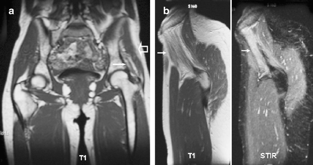
An mri showed possible endometriosis on her iliac muscles and right gluteus muscles.
When pelvic floor muscles are dysfunctional it can lead to a wide range of issues including painful sex intestinal problems like constipation and diarrhea urinary incontinence or problems passing urine and abdominal pain.
An mri is an exam that uses a magnetic field and radio waves to create detailed images of the organs and tissues within your body.
She had no prior diagnosis of endometriosis.
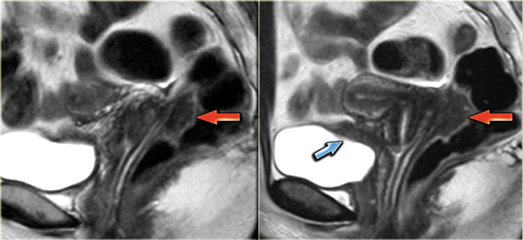
Adhesions can fixate the pelvic organs leading to posterior displacement of uterus and ovaries elevation of the posterior vaginal fornix and angulation of bowel loops.
Mri has greater specificity for the diagnosis of endometriomas than the other non invasive imaging techniques 1 and thus has a role to play in the evaluation of adnexal masses as well as assessing for the response to medical therapy see below potentially eliminating the need for follow up.
Routine and should probably be reserved for known cases of endometriosis rather than for the assessment of pelvic pain.
A good protocol involves.
Pelvic infection is the most frequent gynecologic cause of emergency department visits with the number of such visits approaching 350 000 per year in the united states as many as 70 of the adolescent patients with pelvic inflammatory disease pid are diagnosed in the emergency department and nearly 1 million patients with pid are diagnosed annually in the united states.
Treatment of pelvic.
Evaluation of known endometriosis with mri requires a slightly different protocol to a routine pelvic mri see pelvic mri protocol.
1 it seems women with endometriosis are somewhat more susceptible to having and or developing pfd than those without.
An mri differs from a ct.
9 die can deeply invade into the muscularis propria of the rectosigmoid colon and this deep invasion typically requires surgical resection.
Iv or im buscopan is administered to reduce bowel peristalsis and hence improve image quality.
However her ca 125 levels came back high a possible indicator of endometriosis.
On mri adhesions can be seen as spiculated low to intermediate signal intensity strandings on t1 and t2.
A previous emg spinal mri ct and pet scans came back normal.
Magnetic resonance imaging mri is a noninvasive imaging device that produces strong magnetic field and radio waves that are used to create detailed images of the tissues organs and other areas.
The procedure is generally safe but in rare cases there is a risk of pelvic infection.
For some an mri helps with surgical planning giving your surgeon detailed information about the location and size of endometrial implants.


